EUR 2.6 million in REACT-EU recovery assistance:
New imaging device network advances research [21.04.23]
University of Hohenheim inaugurates five high-performance large-scale research devices / Infrastructure at the Core Facility Hohenheim (CFH) is also available to collaborative partners
The cliché that a picture often says more than 1,000 words holds true for research: A new pool of equipment at the University of Hohenheim in Stuttgart delivers high-resolution three-dimensional visualization of the objects under investigation. Another special feature: The devices can be linked together – so you can view the same detail at different scale levels. On 20 April 2023, the Hohenheim Core Facility put its new research infrastructure into operation with an inauguration ceremony. The equipment was made possible by EU funding in the amount of EUR 2.6 million from the ERDF under the REACT-EU recovery assistance program. From now on, the devices will advance research on the consequences of climate change and species conservation, for example. The BioInterAct research network forms the umbrella for the research projects. In addition to the University of Hohenheim, the University of Tübingen and the Stuttgart Museum of Natural Science are also involved. The new large-scale equipment is available to the working groups of all collaborative partners.
A sustainable agricultural system that defies climate change and preserves biodiversity – that is the long-term goal of the BioInterAct research network. To achieve this, the collaborative partners – the Universities of Hohenheim and Tübingen and the Stuttgart Museum of Natural Science – must keep their research on the ball.
BioInterAct's approach is to gain new insights by visualizing the interactions among plants, insects, and soil. The network under the leadership of Hohenheim systems biologist Prof. Dr. Waltraud Schulze forms an umbrella, so to speak, for numerous research projects.
"Plant development and adaptation to changing environmental conditions as well as insect interactions with plants and soil represent a very complex network. We still have major gaps in our knowledge here," Prof. Dr. Schulze stated. "We want to better understand where external stimuli affect cells and organisms and which structures are important for the interactions."
Coordinated device network provides data basis – and images
This research requires a coordinated network of equipment, which has now been established at the Core Facility Hohenheim (CFH). It consists of various microscopes and an isotope mass spectrometer. "We are very excited about the five new large-scale instruments," said Prof. Dr. Julia Fritz-Steuber. As Vice President for Research, Early Career Researchers, and Transfer, she initiated the application. "The devices allow us to view the same object of study at different scales – from centimeters to nanometers – through different imaging analysis techniques."
She illustrated the advantage with an example: "If you see a bird on a tree and then want to look at it more closely with binoculars, you will have trouble finding the same spot again. We would feel the same way with mismatched microscopes. But with our instrument network, the next scale level zooms directly to the same location on the object under examination." This is particularly interesting for the agricultural sector: “In Hohenheim, we can cover the entire scale from the individual cell to the Agricultural Experiment Station."
Prof. Dr. Fritz-Steuber herself intends to use the new equipment in livestock research. Together with Prof. Dr. Jana Seifert, she studies important bacteria in the rumen stomach of cattle. "Here, another advantage of the instrument network becomes apparent: visualization at the organic, cellular, and molecular levels. Through the imaging process, we can already visually see the composition of the conglomerate of feed substrate and bacteria."
Five high-performance devices – a booster for research
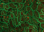
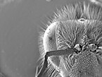
Dr. Wilhelm E. Kincses, head of the Core Facility Hohenheim (CFH), which was also involved in the application, explained the potential uses of the new infrastructure. "With the super-resolution confocal microscope, we can study cellular structures and localize proteins within cells. The confocal laser scanning microscope allows live cell analysis as well as studies of cell structures and whole cells. Finally, the correlative Raman atomic force microscope and the environmental scanning electron microscope allow us to study surface structures of living organisms, among other things."
However, the fifth instrument, a functionally coupled isotope ratio mass spectrometer (IRMS), is important for classifying the results from these imaging techniques. It determines the isotopic composition of different materials: "The isotope ratio can be used to reconstruct the origin of nutrients, create fluxes of substances within plants and determine the interaction of plants with insects," Dr. Kincses said.
The CFH, one University of Hohenheim’s central institutions, operates the large equipment. For this purpose, it is founding its own "Imaging Unit" as a new module. All working groups at the University of Hohenheim, the University of Tübingen, and the Stuttgart Museum of Natural History can now use these services.
Proteins in the cells – response to external stimuli
Prof. Dr. Schulze’s research focuses on the smallest level of investigation, the single cell. That is because small cells can have big effects: the cell is the origin of genetic diversity and influences the entire organism with its adaptation mechanisms. "The proteins and other components in the cells respond to external stimuli, such as extreme weather. From the findings on this, we derive predictions on the next scale level: for example, on the interactions between plants and insects," said Prof. Dr. Schulze.
These interactions are the subject of research by other working groups, which are then able to gain new insights into ecological relationships. The Competence Center Biodiversity and Integrative Taxonomy (KomBioTa), an institution of the University of Hohenheim and the Stuttgart Museum of Natural Science, plays a central role here.
This is followed by greenhouse experiments in the Hohenheim Phytotechnical Center, where researchers investigate the origin of nutrients from the soil under defined conditions. "Ultimately, we want to scale up the findings even further and look at agricultural ecosystems under real conditions, i.e. agricultural systems in Baden-Württemberg," stated Prof. Dr. Schulze. "This is the only way we can get the basis for developing recommendations for action in practice."
BACKGROUND on financial support for the imaging equipment network
In addition to the University of Hohenheim, the University of Tübingen and the Stuttgart Museum of Natural Science are involved in the BioInterAct research network (visualization of plant-soil-insect interactions in climate-stressed agricultural systems). Within the framework of the research network, the new equipment pool was acquired with EU funding from the "European Regional Development Fund" ERDF as part of the Union's response to the Covid-19 pandemic in the amount of EUR 2.6 million.
The invitation to tender for the ERDF program by the Ministry of Science, Research and the Arts of Baden-Württemberg (MWK) was made possible by a top-up from the REACT-EU recovery assistance program. The funds will be used to address the pandemic's aftermath and contribute to a climate-friendly, digital, and stable post-crisis economic recovery.
BACKGROUND: Competence Center Biodiversity and Integrative Taxonomy (KomBioTa)
Species extinction, and in particular the decline of insects, is one of the great challenges of the 21st century. The loss of diversity affects plants, animals, fungi, and microorganisms, the absence of which threatens ecosystem function, and thus important services for humans, such as pollination of plants to fundamental ecosystem services, such as purification of air and water.
To meet these challenges, KomBioTa was established in 2020 at the University of Hohenheim and the Stuttgart Museum of Natural Science with state funding. It bundles numerous working groups at both institutions for joint research and teaching. The state supports the center of the University of Hohenheim and the Stuttgart Museum of Natural Science within the framework of the state initiative "Integrative Taxonomy" with about EUR 1 million annually.
Additional information
Core Facility Hohenheim (CFH): https://cfh.uni-hohenheim.de/en/homepage
ComBioTa: https://kombiota.uni-hohenheim.de/en/english

The isotope ratio mass spectrometer (IRMS) is used to classify the results from the imaging techniques: In particular, the isotope ratio can be used to reconstruct the origin of nutrients from the soil, create fluxes of substances within plants, and quantify the interaction of plants with insects.
Image source: University of Hohenheim / Oliver Reuther
Text: Elsner
Contact for press:
Prof. Dr. Julia Fritz-Steuber, Vice President for Research, Early Career Researchers, and Transfer at the University of Hohenheim,
T +49 711 459-22228, E prorektorat-forschung@uni-hohenheim.de
Prof. Dr. Waltraud Schulze, University of Hohenheim, Department of Plant Systems Biology,
T +49 711 459-24770, E wschulze@uni-hohenheim.de
Dr. Wilhelm E. Kincses, Core Facility Hohenheim (CFH),
T +49 711 459-22671, E wilhelm.kincses@uni-hohenheim.de


